 00389 2 3091 484
Medical services
Electrophysiology
Електрофизиолошка студија
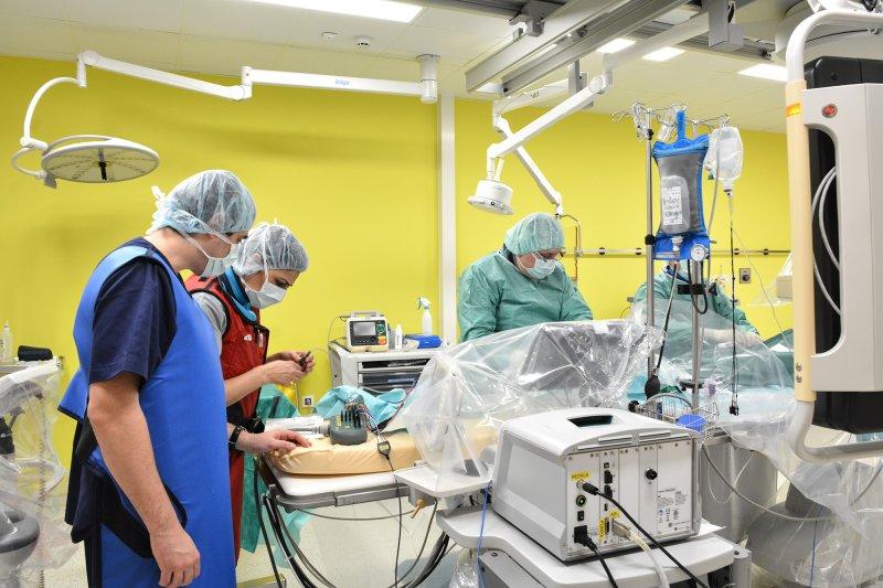
An electrophysiological study is an invasive examination applied to assess, diagnose and treat irregular heart rhythm.
- Reasons for conducting an electrophysiological study
- To treat a documented tachycardia
- To identify the source of the irregular heart rhythm
- To assess the efficiency of applied medications to treat the irregular heart rhythm
- To assess symptoms such as loss of conscience and fast heart rate
- To assess other reasons for the irregular heart rhythm that cannot be assessed using noninvasive methods.
During the electrophysiological study, the physician (a subspecialist for treating heart rhythm disorders – electrophysiologist) examines the electrical system of the heart using several special wires (electrode catheters) placed in the heart, used to record the intracardial electrocardiogram (electrogram). These electrode catheters are inserted through guiding lines placed in the veins of the loins, arms, shoulder, or neck.
After having been placed in the heart, the electrode catheters are used to measure electric signals generated and conducted by the heart. At the same time, electric signals are sent to the heart through these wires, in order to reproduce the abnormal heart rhythm. These basic techniques are used to examine a multitude of heart rhythm disorders, and they are also used to evaluate the effectiveness of the medication treatment of several types of arrythmias.
Once they are positioned in the heart, the electrode catheters are used for measurement of the electrical signals that the heart generates and conducts during its activity. In parallel, electric signals are sent to the heart through the same wires in order to reproduce the abnormal heart rhythm. With these basic techniques, a number of cardiac rhythm disorders are examined, and they are also used for assessment of the efficiency of drug treatment in some arrhythmias.

Heart failure correction Pacemakers or heart resynchronization pacemakers
Heart failure, or as it is also called, a weak heart, is a long-term condition that deteriorates with time. It occurs when the heart muscle is weakened and is not able to pump a sufficient quantity of blood needed to satisfy the requirements of the body to function normally.
In Europe, more than 15 million people die of heart failure annually which makes this condition one of the leading causes of mortality.
Heart failure can be acute, and it usually withdraws after an adequate and timely treatment, but it can also become chronic or end in death.
Failure to recognize, diagnose, and adequately treat heart failure leads to a poor quality of life, frequent hospital treatments, and death.
Cardiac resynchronization therapy (CRT) is a new nonpharmacological method to treat patients with symptoms, under certain conditions, with advanced heart failure – NYHA III-IV, where there usually is a reduced left ventricle ejection fraction <35%, left bundle branch block (LBBB) and an extended QRS complex (>150ms), provided that either dyskinesia or asynchronous motion of the ventricles.
The implantation of this pacemaker stimulates the left and the right ventricle almost simultaneously which, in about 60% of the patients, leads to a recovery of the heart function to a greater or a lesser degree, and sometimes it can lead to a full recovery of the heart as a pump.

Implantable Cardioverter Defibrillators (ICDs)
Implantable Cardioverter Defibrillators (ICDs) – are devices used to treat malignant arrhythmias. The different heart disorders usually lead to an extremely high pulse (180-260 beats/min).
The disruption of the heart rhythm is usually ventricular tachycardia. This is a condition that the patients can tolerate to a certain degree but requires emergency medical assistance. This condition can turn into ventricular fibrillation, a condition when the heart virtually does not perform its pumping function and basically this condition is a reason for sudden cardiac death, unless the patient is resuscitated and treated with electrical shock.
Implantable Cardioverter Defibrillators can recognize these tachyarrhythmias, where special algorithms distinguish these arrythmias from the other tachycardias (acceleration of the heart’s rhythm) and apply an electric treatment wherever necessary.
It terminates them in two ways:
– Through a rapid electric stimulation or the so-called antitachycardia pacing (ATP) which usually normalizes the heart rhythm.
– Where the antitachycardia pacing fails to regulate the heart rhythm, the device applies an electric shock which the patient feels as a shock to the thorax.
– Monomorphic Ventricular Tachycardia
– Ventricular Fibrillation

Pacemaker
A pacemaker is a small electric device, the size of a matchbox, weighing 20-25 grams. It was implanted for the first time in 1958 in Sweden. The basic function of the pacemaker is to provide electric impulses to the heart muscle, thereby causing the muscle to contract.
- Disturbed electric activity of the heart requires the installation of an electro stimulator which will imitate work oof the natural electrical system of the heart.The pacemaker comprises two basic parts: an electrical impulse generator (The “heart” of the pacemaker) and electrodes that carry the impulses from the generator to the heart. Essentially the generator is a place where electric impulses are generated, and they are carried to the heart muscle through thin flexible wires called electrodes.Usually, pacemakers are implanted in patients that have a irregular heartbeat in two cases: slow heartbeat, known as bradycardia, and tachycardia, i.e. a faster heartbeat that cannot be controlled with medication.
The procedure involves your physician injecting a local anesthetic subcutaneously in the wall of the thorax. There the physician will prepare a small pocket from which the electrode(s) (wires) will be inserted into the heart through a nearby vein. The electrodes are placed using an X-Ray machine. After the placement of the electrodes, their function is tested and after obtaining satisfactory test results, they are connected to the pacemaker. The skin is sutured with special sutures that are removed after 7 to 10 days depending on the suture and the suturing.
Before the pacemaker implantation procedure, you discussed with your physician about the possible options and types of pacemakers. After the pacemaker is implanted you will be provided instructions on how to care of the wound. You will be prescribed antibiotics that you will have to continue to take at home. If you need additional information, please do not hesitate to call us whenever you need.
In most cases, the pacemaker implantation procedure is relatively simple and without complications, but there are certain risks that you have to be aware of.
There is a small risk of infection (1 in 400), pneumothorax (lung collapse) (1 in 50), hematoma (1 in 100) or dislocation of the electrodes (1 in 50).
The onset of these procedural risks depends on the individual patient, as well as the existing risk factors and medical conditions.


