Removal of tonsil: When is best to remove them and how it is done in our clinic

Tonsilloadenoidectomy is a surgical procedure for removing palatal tonsils and adenoid vegetations (third tonsil) from the pharynx and the vault of the pharynx (epipharynx). Palatal tonsils and the third tonsil are assimilation of lymph tissue located in the pharynx between the anterior and posterior palatal arches and the vault of the pharynx (epipharynx).

The most common reasons for surgical treatment of tonsils:

  • Frequent recurrent angina (3 in one year)
  • Recurrent peritonsillar abscesses
  • Designated tonsillar hypertrophy (which makes obstructions)
  • Difficult nasal and mouth breathing
  • Snoring during sleep and Sleep apnea
  • If frequent infections affect the incidence of middle ear infections and hearing loss (acute inflammation, secretory otitis media)
  • Carriage, tonsils as a focal point for infections of other organs – (nephritis, rheumatic fever, alopecia) when proven to be the cause of foetor ex ore.

To establish a correct indication for tonsiloadenoidectomy, it is necessary to obtain a good medical history and perform the following tests:

  • ORL examination (oropharyngoscopy, anterior rhinoscopy, otoscopy)
  • Fiber epipharyngoscopy, nasal and throat swab, KKC with CRP with ACO
  • Tympanometry and audiometry
  • Polysomnography (for the diagnosis of sleep apnea)

At the Zan Mitrev Clinic, surgical treatments of tonsils are performed under general endotracheal anaesthesia with a special ULTRACISION-HARMONIC SCALPEL, which reduces the duration of the surgery, as well as the pain and bleeding in the postoperative period.
Schedule examination at: 02/ 30 91 484

Reconstructed aortic valve and creation of 3 new valves in a six-year-old child

Lis Berisha is a 6-year-old child who came for the first time to us for a surgery. He was born with congenital heart defect – aortic stenosis, which is a narrowed opening of the aorta – a large blood vessel coming out of the left ventricle carrying oxygenated blood to the whole body.
Immediately after birth, a systolic murmur was noticed, and due to eating disorders and poor weight gain, the patient was kept in the Prishtina hospital where severe aortic stenosis was identified on an echocardiogram.

– At the age of two months, due to the inability to undergo a surgery, a balloon dilatation of the aortic valve was performed at the University Children’s Clinic in Belgrade, which widened the aortic opening by reducing the pressure gradient. The following regular echocardiographic results showed increased pressure gradient, left ventricular hypertrophy, aortic valve insufficiency with a risk of further heart failure, explained Dr. Jordanka Madjoska, our paediatrician.

It was clear that this child’s condition required surgical treatment that would be a permanent solution without the need for resurgery.
The literature describes three surgery options – replacement of the aortic valve with a mechanical prosthesis, which is a safe and long-lasting solution but requires prevention of thromboembolic complications and prohibition of physical activity or other procedures that cause bleeding; treatment was postponed until the patient comes to a certain age when the biggest prosthesis could be implanted with the greatest risk of re-implantation due to patient’s growth. The second option is to replace the valve with a biological prosthesis that does not last long enough and again requires anticoagulant therapy.

The third option is the Ross procedure – autotransplantation of the pulmonary valve at the aortic position. Of all the possibilities, none was the solution because of the patient’s age.

Our expert team guided by Dr. Zan Mitrev reviewed the case and decided that Lis should be admitted to our clinic and operated. The narrowed aortic part was extended, and Dr. Zan Mitrev designed aortic valves with pericardial matrix material. Immediately after the operation, the follow-ups showed excellent function of the valve and the extended part of the narrowed aorta.

– We provided a permanent solution for the patient because of the material’s ability to grow together with the patient growth and it does not require further surgery, chronic therapy and the patient can practice sports, explained Dr. Madjoska.
The patient had stable vital parameters after surgery and today, one week after the surgery, he is leaving home.
We would define the whole case as an excellent operative and postoperative course of a specific heart defect that was resolved in a way that we can say is new, unique, not yet applied in paediatric cardiac surgery, which will create new protocols in the treatment of aortic stenosis! – concluded the doctor.

Lis and his mother are preparing to leave home today.

– As a mother, I have no words to express the gratitude to the whole team at this clinic, especially to Dr. Zan Mitrev. Thanks for everything you did for my child. All the staff I met at the Zan Mitrev Clinic are extremely professional, with a friendly approach. Now, we go home where our family and especially Lis’ twin brother Malj are expecting us – said Mrs. Berisha.

Dear Lis, be healthy, strong and happy … life is in front of you.

With love,
Zan Mitrev Clinic.

The first prosthesis to replace one part of a leg and the whole joint implanted in our country

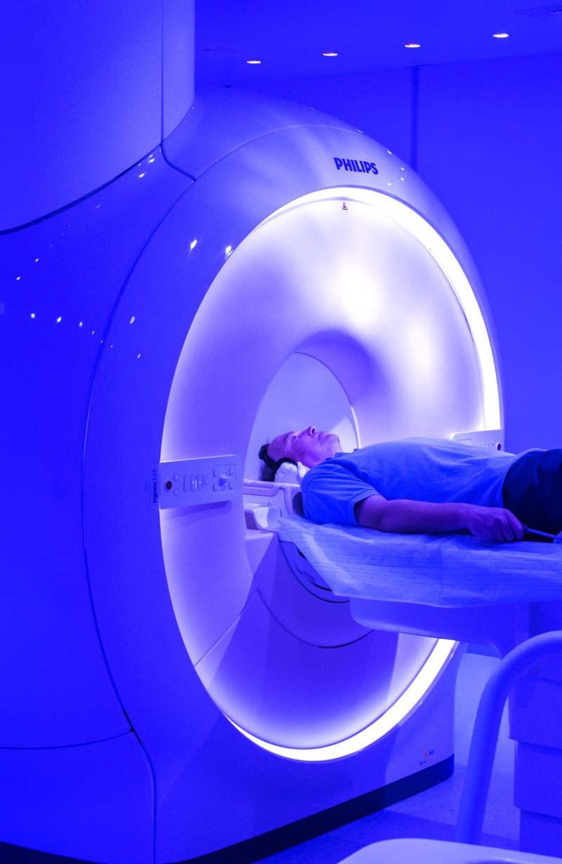
– I had enormous leg pain. I couldn’t walk for months. My whole body was in pain. I had no normal life for several years. My whole life has gotten worse … I was in agony. Despite the two surgeries performed in public hospitals, the tumour I had in my knee returned. I was desperate and did not know if there was any salvation for my leg, which besides being unable to use, it caused terrible pain. I couldn’t sleep, I couldn’t eat, I couldn’t live … – this is how the young woman from Veles, who left our clinic waking on both feet, started her life story despite the difficult situation she was in when admitted to our clinic.

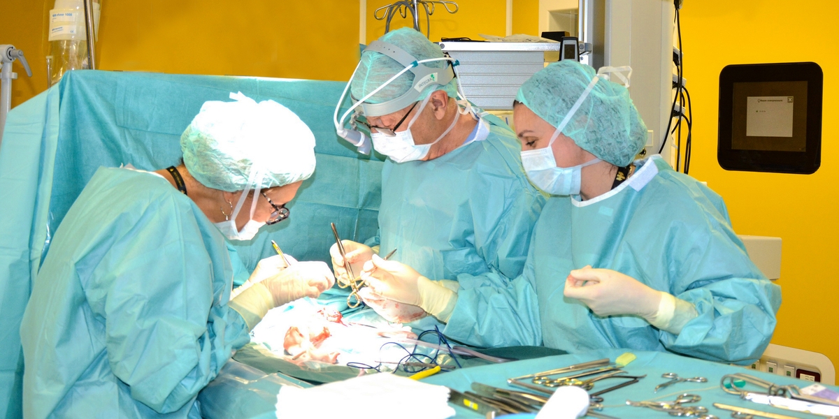
The Zan Mitrev Clinic orthopaedics team, in this case guided by the orthopaedic surgeon Dr. Sasho Mladenovski, had a real challenge to save a young life and enable limb preservation, i.e., the left leg and a normal life in future. With the long-lasting complicated surgery, a large portion of the lower leg bone was removed, and for the first time in our country, a successful LPS rotation hinge (Limb preservation system) was implanted, which implies a modular implant system that could preserve the limb completely and establish its normal function.

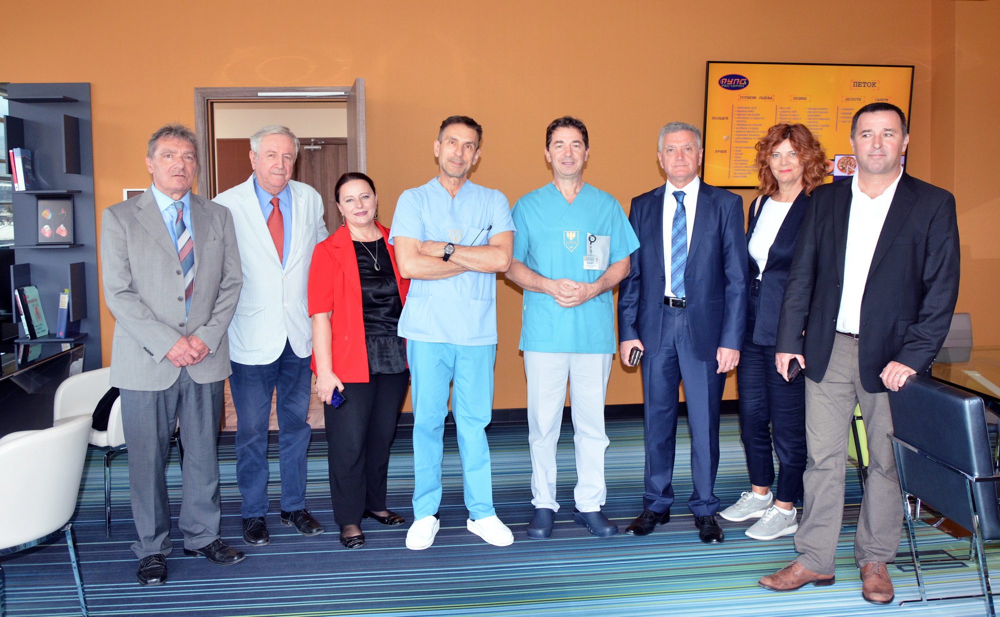
Dr. Mladenovski explained how the patient was admitted at the Zan Mitrev Clinic, what was her diagnosis and how she was treated:

– The patient was diagnosed with an aggressive giant-cell tumour of the lower leg bone. She was twice operated in another public health facility, but after both surgeries, the same tumour was growing progressively, destroying the lower leg bone and upper joint at the height of 12 cm.

– The patient came to us in severe pain and unable to walk, referred by the Health Fund to private hospitals including ours, asking if this patient could be cared for and treated. With more than 2000 surgeries performed, with primary, revision and tumour primary endoprostheses of knees, hips and shoulders implanted, our orthopaedic team accepted her and agreed t be treated. So, the patient stayed here and received treatment – explained the doctor Mladenovski.

He emphasized that with this operation, a LPS revision rotation hinge cementless knee prosthesis has been implanted for the first time in our country.

– It is a modular system used in major bone defects caused by destructive neoplastic processes of the bones and joints, in people with poor healing of intraarticular fractures and severe osteoporosis, in patients requiring recurrent revision surgeries with pre-placed primary or revision implants on the large joints. This patient had a radical resection of the tumour itself, which means that more than a third of the lower leg bone is missing and replaced by a system that completely replaces the lost bone structure. In addition to the bone structure, this system complements the lack of all the necessary ligaments of the knee joint itself, which were sacrificed in the procedure for removal of the tumour – explained Dr. Mladenovski.

This complex system was successfully implanted. After the operation, the patient stayed in the hospital for another 7 days and gradually started and early physical therapy, such as cryo-therapy, gradual standing up and slight leg loading, because a period of 6 to 8 is needed for a complete adhesion of the cementless bodies placed inside the bones.

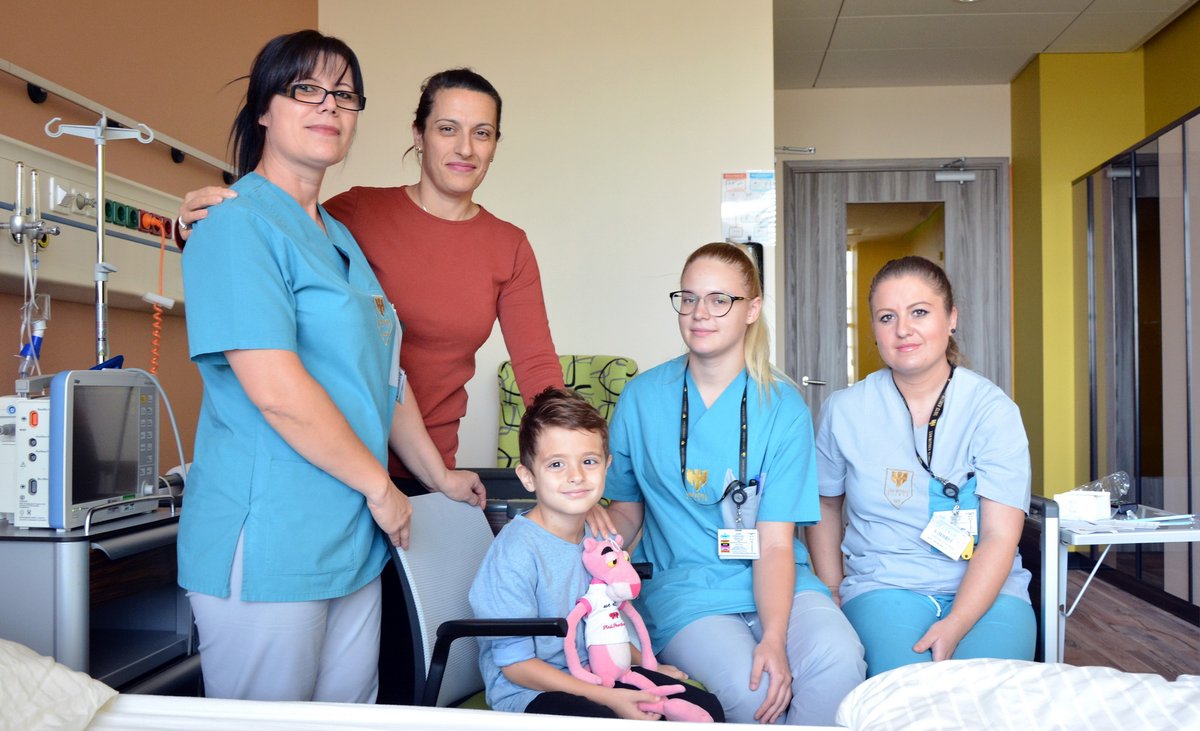
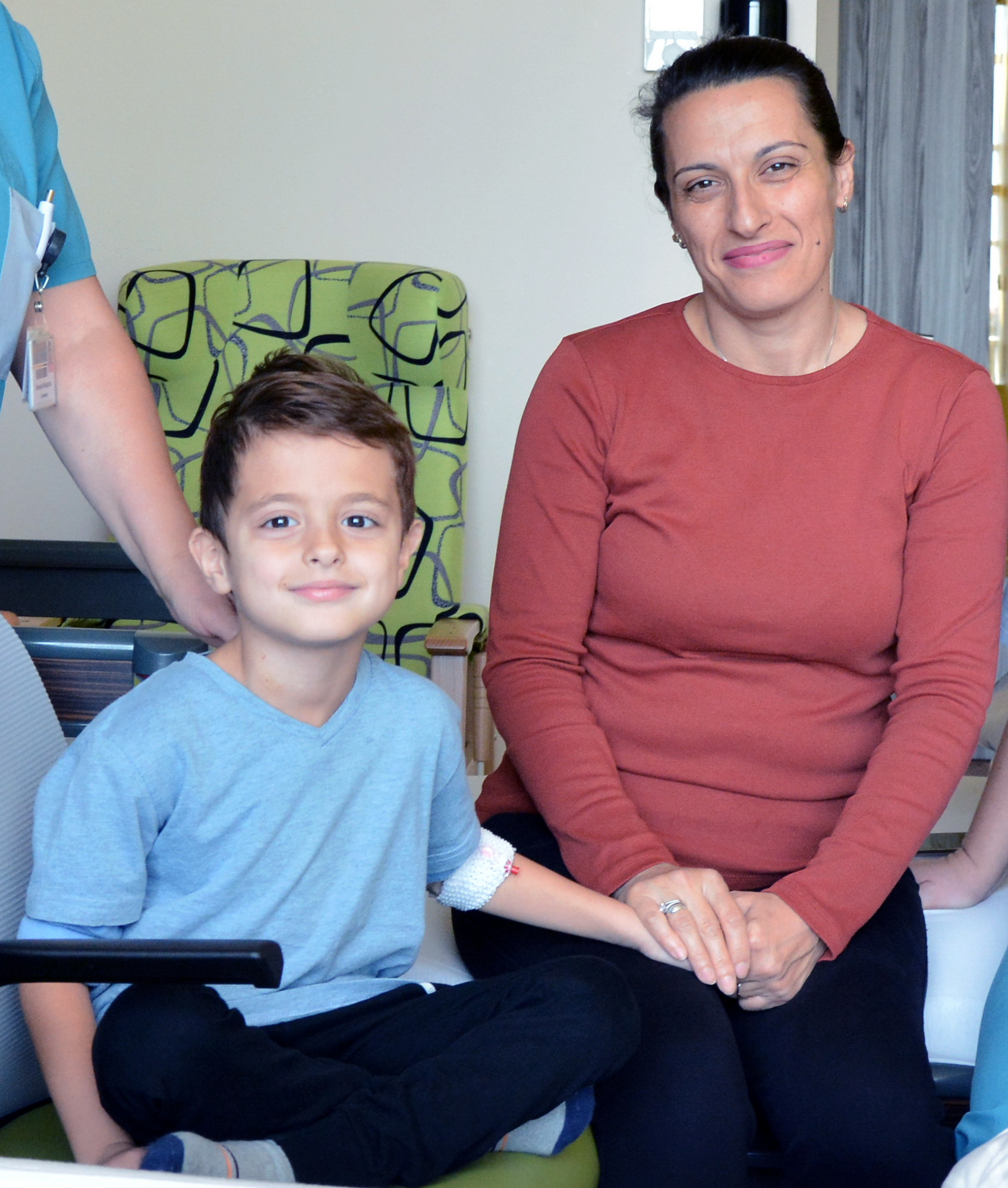
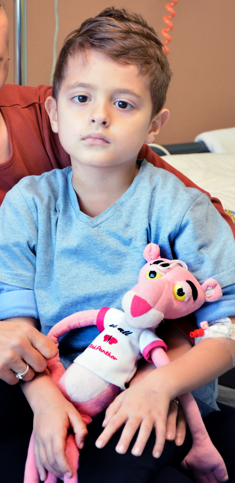
– The patient had a great recovery and is currently undergoing physical therapy. She started to make full steps with the operated leg, which means she started daily activities, and I I am happy to say that the surgery is successfully completed with the best possible outcome available for this type of surgery – concludes Doctor Mladenovski.

At the first follow-up, the patient felt great walking with both legs. Soon, she will e able to walk without the crutch and return fully to daily life.


The Shibenik Mayor Zheljko Buric meets with Dr. Zhan Mitrev

Organized by the NGO “Croatian-Macedonian Tangent”, today the Mayor of Shibenik Zheljko Buriс and his associates Mirjana Zhuric – Head of Social Activities, and Dino Karadzole – Director of the Tourist Community of the City of Shibenik, visited the Zhan Mitrev Clinic. They met the first man of the clinic, Dr. Zhan Mitrev and discussed several topics related to both medicine and the organizational segments of a company.

– This visit to Zhan Mitrev Clinic is very interesting from several aspects. Since I am a doctor, ophthalmologist, and I have been the manager of the Shibenik Hospital for 14 years, and now the Mayor of Shibenik since 2013, we have a lot of areas to discuss, which are of my interest. I learned about this hospital by reading about it and watching videos and reports. It is very important for me to see how a modern and impeccably organized private clinic with such an extensive program of medical services operates in order to replicate it on the public health in Croatia, which has organizational problems. Once I got in your hospital, I immediately saw the difference, from the investment amount to the way it is organized. What particularly impressed me was the control of intra-hospital infections, which is a huge problem in almost all public health units – said Mayor Buric.

He announced a possible collaboration in the field of medical tourism, primarily in the form of knowledge acquisition and replication of the Zhan Mitrev Clinic system in his municipality and state.

After the visit to our clinic, our guest from Croatia and his delegation will meet with persons and institutions from the public and political life of the Macedonian capital. In addition to the meeting with Skopje Mayor Petar Shilegov, the delegation of the City of Shibenik will also visit the Macedonian Parliament, as well as government agencies for emigration and tourism promotion.

A meeting with representatives of the Croat Community in Macedonia is also planned.

Two tumours were removed, one of a large size, from a patient who had been referred from hospital to hospital for years

In our clinic, one of the many characteristic cases that have been observed lately is the case of a 56-year old woman, who has experienced a real trauma and struggle for her life in other clinics where she was previously treated without solving her life-threatening problem. She had a history of liver hemangioma proven 5 years ago. The last MSCT control (multi-slice computed tomography) before coming to Zan Mitrev Clinic was in 2018 after which she reported to our clinic due to dissatisfaction with public health services.

– The patient’s weight is 95 kg, her height is 164 cm, and body mass index (BMI) of 35.33 kg/m2. The main complaints of the patient were pain in the right rib arch and in the right shoulder. With the control MSCT, we found that the hemangioma occupied almost the entire right lobe of the liver (about twenty centimeters in diameter and several kilograms of weight), with a concurrent and smaller hemangioma in the left lobe. Compared to the previous MSCT finding, the hemangioma was now increased by almost 50%. At the level of the hepatic veins, compression and pathological anastomosis with the inferior vena cava have been established. Reconstruction of the abdominal artery showed, as expected, that there was no pathological communication between the right hepatic artery and the hemangioma. Because of the size of the change, surgical removing of the two liver changes was advised – explained Dr. Milcho Panovski, an abdominal surgeon and subspecialist at our clinic, who later performed this complicated surgery with the team.

After the immediate preparation, the same team of the Zan Mitrev Clinic performed the surgery. The patient was prepared and guided in accordance with the ERAS (Enhanced Recovery After Surgery) protocol. The team that performed the surgery explained that because of the size of the hemangioma, the operational field was significantly limited, and the operation aggravated. During the surgery, due to expected bleeding, IOS (intraoperative blood salvage) was performed, i.e, a medical procedure that involves recovering of the blood lost during surgery and re-inserting it into the patient. This procedure is a major form of self-transfusion.

– We managed to safely remove the hemangioma from the right lobe and the other one from the left lobe. The total operation time was 210 min. The surgery and post-surgical flow were normal – added Dr. Panovski.

After surgery, the patient is in stable health condition, recovering at home.

“They told me I have a tumour, which must be removed, but due to the summer holidays there is no one to do it”


In our clinic, often our doctors respond quickly, decisively and correctly. Fighting for the life of the patient is a priority that in the race against time becomes the sole goal of all of us. This fight uses all available means, both technical and human, regardless of time and place. Exactly such a fight, which is very common in our clinic, happened for the life of one of our citizens who (imagine !) was not admitted by one of the state hospitals, i.e., was not urgently operated on, though his condition required urgent surgery, “because of the start of the summer holidays”.

  • Although I was in pain and moving very hard, I was told in the state hospital that there was no one to operate me. They did examinations and told me I had a tumour, which must be surgically removed as soon as possible, but due to the start of the summer holidays there was no one to do it. It was clear to me that I had to look for salvation elsewhere, after which my family decided to go to the Zhan Mitrev Clinic. Here I was operated on, and I want to express enormous gratitude to the whole team that treated me – said the patient, recalling everything he had gone through.

His story at the Zhan Mitrev Clinic begins when he was admitted and immediately hospitalized. After performing all tests, surgery was scheduled.

– It has been found that the 71-year old patient (56 kg heavy, 162 cm high, BMI 21.34 kg/m2) had neurofibrosarcoma on the left side in the retroperitoneum. Clinical examination revealed a large palpable tumour in the abdomen that clearly protrudes. There was also a visible wound from a previous biopsy. Laboratory tests showed uncontrolled glycemia, anaemia, thrombocytosis (Plt 406.1 x 10³/µL), malnutrition (Albumin 22.0 g / l). The MSCT showed that the tumour infiltrates the left kidney and its blood vessels, pushes the colon and has a doubtful relation to the mesenteric blood vessels of the small intestine. Simultaneously, it pushes/infiltrates the pancreas and compresses the lienal vein. Due to anaemia and malnutrition, the preparation for surgery was made according to the ERAS (Enhanced Recovery After Surgery) protocol – informed Prof. Milcho Panovskim MD, Abdominal Surgeon, Subspecialist in the Zhan Mitrev Clinic.

The patient underwent combined (general and epidural) anaesthesia. Intraoperatively, it has been determined that there are metastatic changes in the omentum and radical procedure was possible.

Our combined oncology team (urologist, abdominal surgeon) successfully completed the surgery. The large tumour, 27 x17 x 14 cm, along with the omentum, was removed. Removal of a body part and the tail of the pancreas, along with the spleen, has also been done. Due to tumour infiltration in the colon blood vessels, a partial resection of the colon with termino-terminal anastomosis was performed.

  • Total surgery duration – 260 minutes.
  • Post-operative replacement therapy for pancreatic exocrine and endocrine function, prophylaxis for thrombocytosis, and the patient was guided according to the ERAS protocol
  • The post-operative course is normal, with one-day psychogenic disorientation present.

Zan Mitrev Clinic participant of the 6th Congress of the Macedonian Society of Cardiology

The 6th Congress of the Macedonian Society of Cardiology with international participation was held from October 3rd to October 6th in Ohrid. The Congress was for cardiologists, cardiology fellows and other professionals working in the field of cardiology, who participated in different topics of the cardiovascular medicine.

The Zan Mitrev Clinic had also its representatives at the congress, of course with its first lecturer Dr. Zan Mitrev followed by lectures of Dr. Ivan Milev, Dr. Predrag Milojevic and Dr. Tanja Andjusheva.

One of the main topics at the Congress were cardiovascular imaging techniques, which are a very important segment in the diagnosis, prognosis, treatment decision and monitoring of cardiovascular diseases. Coronary and structural myocardial diseases treated with percutaneous interventions, cardiac arrhythmias and peripheral artery disease were the topics that were also the focus of the National Congress of the Macedonian Society of Cardiology.

Of course, coronary artery disease, heart failure, cardiomyopathy, preventive cardiology, pharmacotherapy, cardiac surgery, pediatric cardiology, and other cardiovascular diseases were also presented and discussed at the Congress.

The congress was composed of plenary sessions with national and international experts, symposia, oral and poster presentations in the field of modern cardiovascular medicine.

During the Congress, the First Balkan Congress of Nurses and Technicians was also held, where our clinic took a prominent place with its representatives.

This Congress is an opportunity to exchange experiences on the latest scientific and professional achievements in cardiology and to listen and meet colleagues from home and abroad.

What is Cardiac Magnetic Resonance Imaging and how it is performed

Cardiac Magnetic Resonance Imaging (MRI) is a non-invasive diagnostic method that uses a magnetic field and radiofrequency to produce a detailed image of the heart, blood vessels and surrounding organs.

* Cardiac Magnetic Resonance Imaging is used for:

  • Checking for tissue damage of myocardial infarction, what the damage is and whether it is permanent (myocardial viability)
  • Accurate assessment of heart failure, potential causes of heart failure
  • Evaluation and diagnostics of cardiomyopathies (dilated, hypertrophic, restrictive amylodosis)
  • Diagnostics of myocarditis (inflammation of the heart muscle)
  • Valve defects
  • Evaluation of pericardium (lining around the heart)
  • Assessment of congenital heart anomalies

Cardiac MRI is a safe and painless diagnostic procedure. People with any metal devices in and on the body must not undergo this procedure without confirmation that the device is MRI safe.

Such devices include:

  • Cardiac pacemakers, implantable cardiac defibrillators (ICDs) and resynchronization therapy devices (CRTs)
  • Inner ear implants
  • Implanted infusion pumps, neuro-muscle stimulators, intrauterine contraceptive devices
  • Brain aneurysm clips
  • Some dental implants
  • Any metal body parts

Prior to the examination, the patient should report on possible pregnancy, having tattoos, stents, valve prosthesis, heart surgery or kidney disease.

What happens during the examination?

  • The examination usually takes 30-60 minutes
  • The patient is lying on a comfortable moving table with headphones listening to the technologist’s instructions
  • Patient cooperation is required with regard to breathing, exhaling and holding breath for a few seconds
  • ECG and oxygen saturation are monitored all the time, and the patient is free to speak to the technologist during the examination
  • If contrast application is needed, intravenous cannula in the arm is planned prior to examination.

After the examination, immediate return to normal activities is possible. There is no limit as there is no ionizing radiation. The examination results are usually obtained within 2-3 business days.