
 00389 2 3091 484
00389 2 3091 484

What is Cardiac Magnetic Resonance Imaging and how it is performed
October 3, 2019
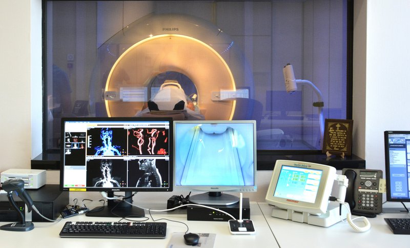
Cardiac Magnetic Resonance Imaging (MRI) is a non-invasive diagnostic method that uses a magnetic field and radiofrequency to produce a detailed image of the heart, blood vessels and surrounding organs.
* Cardiac Magnetic Resonance Imaging is used for:
- Checking for tissue damage of myocardial infarction, what the damage is and whether it is permanent (myocardial viability)
- Accurate assessment of heart failure, potential causes of heart failure
- Evaluation and diagnostics of cardiomyopathies (dilated, hypertrophic, restrictive amylodosis)
- Diagnostics of myocarditis (inflammation of the heart muscle)
- Valve defects
- Evaluation of pericardium (lining around the heart)
- Assessment of congenital heart anomalies
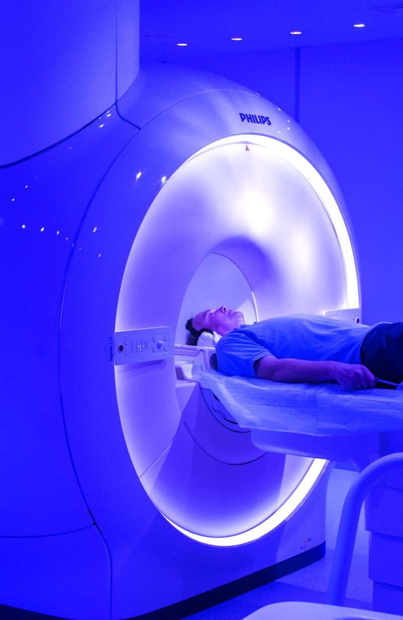
Cardiac MRI is a safe and painless diagnostic procedure. People with any metal devices in and on the body must not undergo this procedure without confirmation that the device is MRI safe.
Such devices include:
- Cardiac pacemakers, implantable cardiac defibrillators (ICDs) and resynchronization therapy devices (CRTs)
- Inner ear implants
- Implanted infusion pumps, neuro-muscle stimulators, intrauterine contraceptive devices
- Brain aneurysm clips
- Some dental implants
- Any metal body parts
Prior to the examination, the patient should report on possible pregnancy, having tattoos, stents, valve prosthesis, heart surgery or kidney disease.
What happens during the examination?
- The examination usually takes 30-60 minutes
- The patient is lying on a comfortable moving table with headphones listening to the technologist’s instructions
- Patient cooperation is required with regard to breathing, exhaling and holding breath for a few seconds
- ECG and oxygen saturation are monitored all the time, and the patient is free to speak to the technologist during the examination
- If contrast application is needed, intravenous cannula in the arm is planned prior to examination.
After the examination, immediate return to normal activities is possible. There is no limit as there is no ionizing radiation. The examination results are usually obtained within 2-3 business days.


Related News
17.06.2025
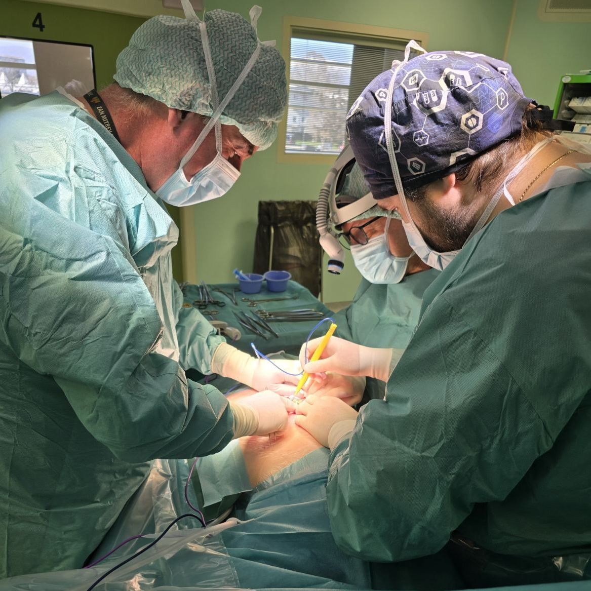
Quick and simple solution to cryptorchidism or undescended testicle condition
Cryptorchidism is a condition in which the testicle has not descended into the scrotum and is instead located hidden inside…
View More
07.05.2021
FROM TOMORROW: COVID-19 TESTING AT THE OHRID ST. PAUL THE APOSTLE AIRPORT
With the beginning of the summer flight schedule at the Ohrid St. Paul the Apostle Airport and in accordance with…
View More27.01.2021
TAV Macedonia and Zan Mitrev Clinic with solution to the new travel restrictions: Rapid antigen tests at Skopje International Airport
In order to respond to the latest travel restrictions imposed due to COVID-19 by EU/ Schengen countries, TAV Macedonia together…
View More